
We take care of diseases of the digestive system
As gastroenterologists, we primarily treat diseases of the digestive system, i.e., the oesophagus, stomach, small and large intestine, liver, and pancreas. We prioritize making your experience as comfortable and stress-free as possible during consultations, examinations, and treatments. Our practice meets the highest professional, technical, and hygienic standards to ensure the best possible care.

Do you need a screening colonoscopy?
If you are over 50, symptom-free, and considering a screening colonoscopy, you can schedule the necessary preliminary consultation by booking an appointment here.
For all other appointments, please call us at +497071 5005
Overview of our examination techniques
We provide care for conditions of the digestive system, particularly involving the esophagus, stomach, small and large intestines, liver, and pancreas.
GASTROSCOPY
A gastroscopy is usually used to clarify complaints in the upper abdomen or chronic diarrhea. Certain diseases such as a reflux of gastric acid into the esophagus, chronic gastritis (e.g., due to infection with Helicobacter pylori), stomach or duodenal ulcers or coeliac disease can be detected.
Examination procedure
The examination uses a thin, soft tube (endoscope), which is inserted through the mouth and advanced through the esophagus and stomach into the first part of the small intestine, known as the duodenum. The endoscope contains glass fibers to transmit light, and its tip can be maneuvered in all directions. A tiny camera at the end of the scope, similar to those found on today’s smartphones, can be used to view and photograph the mucous membrane of the organs. In most cases, taking small tissue samples, so-called biopsies, is also necessary. This is done using small forceps inserted through the endoscope and operated from the outside.
Since endoscope insertion usually is uncomfortable, the procedure is typically performed under sedation (a short period of artificial sleep). After sedation, you are legally restricted from driving or operating machinery until the following day, so it is advisable to arrange transportation home.
Only an empty stomach can be examined properly and safely; therefore, you should not eat anything from 10 p.m. the day before and not drink anything for 4 hours before the examination. The exception is morning medication with a small sip of water.
COLONOSCOPY
A colonoscopy is most commonly performed as a preventive measure for colon cancer. During the procedure, benign precursors of colon cancer, known as polyps or adenomas, are removed in about one-fifth to one-third of the examinations. This prevents this dangerous disease in approximately 90% of cases. Therefore, a colonoscopy is recommended for everyone from the age of 50, every 10 years. In particular (familial) situations, it may be advised earlier and more frequently. Additionally, a colonoscopy can be used to investigate chronic diarrhea and intestinal bleeding. For example, it can help diagnose chronic inflammatory bowel diseases such as Crohn’s disease and ulcerative colitis, or vascular changes in the intestinal mucosa, which can sometimes also be treated during the procedure.
Examination procedure
The examination is carried out using a soft, finger-thick tube that is inserted through the anus and advanced to the beginning of the large intestine and often a short distance into the end of the small intestine. Light is directed into these organs through fiber optics in the endoscope. Its tip can be angled in all directions by turning wheels at the outer end of the endoscope. At the tip, there is also a tiny camera, similar to those found in modern smartphones, which is used to view and photograph the mucosa of the organs. Removal of polyps or the collection of small tissue samples, known as biopsies, is performed using special instruments being advanced through the endoscope and operated from outside.
Because pushing forward the colonoscope is uncomfortable or painful for most people, the procedure is usually performed under sedation. After such sedation, you are legally not allowed to actively participate in road traffic until the next day. Therefore, it is best to arrange for someone to pick you up after the examination.
Only an empty bowel can be examined properly and safely. Depending on the time of the examination, specific preparation is required, starting in the afternoon or evening of the previous day. You can find more information about this preparation here.
In a personal consultation scheduled before the examination, we will explain all the steps in detail. We will also be happy to answer any questions about the procedure.
Important information on preparation
CAPSULE ENDOSCOPY
In addition to endoscopy of the gastrointestinal tract using an endoscope, there is also the option of swallowing a tiny camera capsule. An capsule endoscope is a tube-shaped device with a built-in light source and miniature camera, which is usually inserted into the body via the mouth or anus during a short period of artificial sleep. Such a camera capsule – about the size of a vitamin tablet – is also equipped with a camera and a flashlight, but also with a battery and a radio transmitter so that it can send up to 6 images per second from inside the body to the outside on its way through the digestive tract. These, together with the position of the capsule in the body during the examination, are stored on a hard disk by a small receiver device worn on the body.
After the examination, the images (up to 70,000) can be viewed and analyzed like a video. The capsule is a single-use item disposed of via the toilet.
Capsule endoscopy provides a way to examine the small intestine, which is otherwise difficult to access. The large intestine can also be examined with a specially programmed capsule.
Preparation and Cost coverage
Before undergoing such an examination, it is necessary to confirm—using a CT/MRI scan or a special test capsule—that the capsule can pass through the entire gastrointestinal tract without any problems. As with a traditional colonoscopy, the bowel must be cleaned using appropriate laxative measures.
Capsule endoscopy is typically covered by health insurance for certain diagnostic purposes. In other cases, the insurance company’s prior approval of cost coverage should be obtained.
Small intestine capsule – capsule enteroscopy
Due to its location between the stomach and large intestine and its length of approximately 5 meters, the small intestine is not accessible with conventional endoscopes. Examination with a video capsule (capsule enteroscopy) is particularly suitable for visualizing the small intestine’s mucous lining along its entire length.
This method is most often used for cases of unexplained blood loss when the source of bleeding cannot be identified with a gastroscopy or colonoscopy. This situation can occur, for example, during anticoagulant (“blood-thinning”) treatment.
Unexplained abdominal pain and diarrhea that cannot be clarified using other methods may indicate an inflammatory bowel disease. Capsule enteroscopy can help determine the extent of potential small intestine involvement.
Large Intestine capsule – capsule colonoscopy
Examining the large intestine using a video capsule (capsule colonoscopy) can be an alternative to conventional colonoscopy if the latter is either unfeasible or not desired. Comparative studies show that, with optimal preparation and execution, the diagnostic accuracy is similar to that of conventional colonoscopy. As with a colonoscopy, bowel preparation through cleansing is required.
The advantages arethe practical lack of risk of injury andthat no sedation is required for the examination, so the ability to work is theoretically given, and the ability to drive a motor vehicle and conduct business is not restricted.The main disadvantages are the lack of intervention options, i.e. no mucosal samples can be taken and no polyps removed in the event of abnormalities. In such a case, a conventional colonoscopy with further preparation is usually required.
Important information on preparation
ULTRASOUND
The following can be examined using ultrasound:
- the abdominal organs (liver, gallbladder and bile ducts, pancreas, spleen, kidneys, intestines, urinary bladder, prostate, uterus, ovaries including the associated blood vessels)
- the chest (e.g., to detect pneumonia, pleurisy, or a rib fracture)
- the thyroid gland
- the blood vessels in the neck, legs (e.g., to detect thrombosis) and arms
Contrast-Enhanced Ultrasound
Contrast-enhanced ultrasound is a technique in which a contrast agent (more specifically, an echo signal enhancer) is injected intravenously via an infusion needle during the procedure. Over the following minutes, blood flow can be monitored and recorded in real-time, allowing for dynamic visualization. This method is particularly useful for assessing whether liver lesions are benign or malignant.
The advantage of this method over radiological methods such as computer tomography (CT) or magnetic resonance imaging (MRI) is that it is well tolerated: on the one hand, the contrast medium, which cannot cause any side effects on the kidneys or thyroid gland, and on the other hand the ultrasound, which has no damaging effect on the tissue. Furthermore, additional information can be obtained through the uninterrupted recording of the blood flow behavior.
The main disadvantage is that several sites in an organ or in the body cannot be examined simultaneously, but at best one after the other, and that some sites in the body cannot be visualized with sufficient quality using ultrasound.
Preparation
You will be informed about the contrast agent administration at least a day before the examination. Therefore, two appointments are generally required. You can review the consent form in advance by clicking the link below.
Your way to us
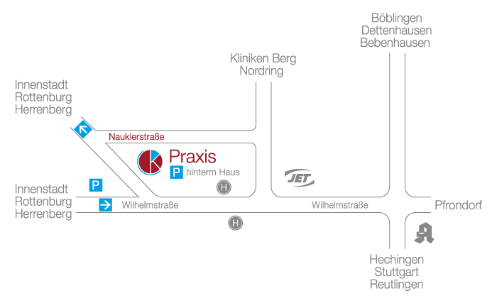
Address
MVZ für Diagnostik, Prävention, Onkologie und Gastroenterologie Tübingen GmbH
Dr. med. Johannes von Keller
Nauklerstraße 50
72074 Tübingen
Telephone: 07071 50 05
Fax number: 07071 50 06
gastro@mvz-tuebingen.de
We operate purely on an appointment basis. You are welcome to make an appointment for a consultation or examination by calling +497071 5005